Students can Download Chapter 6 Breathing and Exchange of Gases Notes, Plus One Zoology Notes helps you to revise the complete Kerala State Syllabus and score more marks in your examinations.
Kerala Plus One Zoology Notes Chapter 6 Breathing and Exchange of Gases
What is respiration?
The process of exchange of O2 from the atmosphere with CO2 produced by the cells is called breathing, commonly known as respiration.
![]()
RESPIRATORY ORGANS:
| 1. Lower invertebrates like sponges, coelenterates, flatworms, etc. exchange O2 with CO2 by simple diffusion over their entire body surface. 2. Earthworms use their moist cuticle and insects have a network of tubes to transport atmospheric air within the body. 3. Gills are used by most of the aquatic arthropods and molluscs whereas lungs are used by the terrestrial forms for the exchange of gases. 4. Fishes use gills whereas reptiles, birds and mammals respire through lungs. 5. Frogs can respire through their moist skin also. 6. Mammals have a well developed respiratory system. |
Human Respiratory System:
The nostrils leads to a nasal chamber through the nasal passage. The nasal chamber opens into nasopharynx the common passage for food and air. Nasopharynx opens through glottis of the larynx region into the trachea.
Sound box in respiratory system:
Larynx is a cartilaginous box which helps in sound production called as the sound box. Glottis is covered by a thin elastic cartilaginous flap called epiglottis to prevent the entry of food into the larynx. Trachea is a straight tube which divides into a right and left primary bronchi.
Each bronchi undergoes repeated divisions to form the secondary and tertiary bronchi and bronchioles ending up in very thin terminal bronchioles.
Each terminal bronchiole gives rise to vascularised bag-like structures called alveoli. The branching network of bronchi, bronchioles and alveoli comprise the lungs.
The two lungs which are covered by a double layered pleura, with pleural fluid between them. It reduces friction on the lung surface. The conducting part transports the atmospheric air to the alveoli. Exchange part is the site of diffusion of O2 and CO2 between blood and atmospheric air.

Where is lung fitted in human body?
The lungs are situated in the thoracic chamber which is formed dorsally by the vertebral column, ventrally by the sternum, laterally by the ribs and on the lower side by the dome-shaped diaphragm. The anatomical setup of lungs in thorax is the arrangement essential for breathing, directly alterthe pulmonary volume.
Respiration involves the following steps:
- Breathing or pulmonary ventilation by which atmospheric air is drawn in and CO2 rich alveolar air is released out.
- Diffusion of gases (O2 and CO2) across alveolar membrane.
- Transport of gases by the blood.
- Diffusion of O2 and.CO2 between blood and tissues.
- Utilisation of O2 by the cells for catabolic reactions and resultant release of CO2
![]()
MECHANISM OF BREATHING:
Breathing involves two stages:
- Inspiration during which atmospheric air is drawn in and
- Expiration by which the alveolar air is released out.

How does increase or decrease of pulmonary volume occur?
Inspiration is initiated by the contraction of diaphragm which increases the volume of thoracic chamber .The contraction of external inter-costal muscles lifts up the ribs and the sternum causing an increase in the volume of the thoracic chamber. It increase the pulmonary volume.
An increase in pulmonary volume decreases the intra-pulmonary pressure to less than the atmospheric pressure which forces the airfrom outside to move into the lungs,i.e., inspiration Relaxation of the diaphragm and the inter-costal muscles returns the diaphragm and sternum to their normal positions and reduce the thoracic volume.
It reduce the pulmonary volume. This leads to an increase in intra-pulmonary pressure to slightly above the atmospheric pressure causing the expulsion of airfrom the lungs, i.e., expiration.
Instrument used for measuring breathing movements:
A healthy human breathes 12 – 16 times/minute. The volume of air involved in breathing movements can be estimated by using a spirometer.
![]()
Respiratory Volumes and Capacities:
| Tidal Volume (TV): Volume of air inspired or expired during a normal respiration. It is approx. 500 mL, i.e. a healthy man can inspire or expire approximately 6000 to 8000 mL of air per minute. |
| Inspiratory Reserve Volume (IRV): Additional volume of air, a person can inspire by a forcible inspiration. This averages 2500 mL to 3000 mL. |
| Expiratory Reserve Volume (ERV): Additional volume of air, a person can expire by a forcible expiration. This averages 1000 mL to 1100 mL. |
| Residual Volume (RV): Volume of air remaining in the lungs even after a forcible expiration. This average 1100 mL to 1200 mL. |
| Expiration Capacity (EC): Total volume of air a person can expire after a normal inspiration. This includes tidal volume and expiration reserve volume (TV + ERV). |
| Inspiratory Capacity (IC): Total volume of air a person can inspire after a normal expiration. This includes tidal volume and inspiratory reserve volume (TV+IRV). |
| Functional Residual Capacity (FRC): Volume of air that will remain in the lungs after a normal expiration. This includes ERV + RV. |
| Vital Capacity (VC): The maximum volume of air a person can breathe in after a forced expiration. This includes ERV, TV and IRV or the maximum volume of air a person can breathe out after a forced inspiration. |
| Total Lung Capacity: Total volume of air accommodated in the lungs at the end of a forced inspiration. This includes RV, ERV, TV and IRV or vital capacity + residual volume. |
EXCHANGE OF GASES:
Alveoli are the sites of exchange of gases. O2 and CO2 are exchanged in these sites by simple diffusion.
Rate of diffusion:
Solubility of the gases and the thickness of the membranes are the important factors that affect the rate of diffusion.

Partial pressure of gases:
Pressure contributed by an individual gas in a mixture of gases is called partial pressure and is represented as pO2 for oxygen and pCO2 for carbon dioxide. As the solubility of CO2 is 20 – 25 times higher than that of O2, the amount of CO2 that can diffuse through the diffusion membrane.

![]()
As the solubility of CO2 is 20 – 25 times higher than that of O2 the amount of CO2 that can diffuse through the diffusion membrane.

The diffusion membrane is made up of three major layers such as
| 1. The thin squamous epithelium of alveoli, 2. The endothelium of alveolar capillaries and 3. The basement substance in between them. |
Its total thickness is much less than a millimetre.
TRANSPORT OF GASES:
Blood is the medium of transport for 02 and C02.
- About 97 per cent of O2 is transported by RBCs in the blood.
- The remaining 3 per cent of O2 is carried in a dissolved state through the plasma.
- Nearly 20 – 25 per cent of CO2 is transported by RBCs whereas 70 per cent of it is carried as bicarbonate.
- About 7 per cent of CO2 is carried in a dissolved state through plasma.
![]()
Transport of Oxygen:
O2 bind with haemoglobin to form oxyhaemoglobin. Each haemoglobin molecule can carry a maximum of four molecules of O2. Binding of oxygen with haemoglobin is related to partial pressure of O2.
| A sigmoid curve is obtained when percentage saturation of haemoglobin with O2 is plotted against the pO2. This curve is called the Oxygen dissociation curve. |
Oxygen dissociation curve is useful in studying the effect of factors like pCO2, H+ concentration, etc., on binding of O2 with haemoglobin.
| In the alveoli, high pO2, low pCO2, lesser H+ concentration and lower temperature are all favourable for the formation of oxyhaemoglobin. |
| In the tissues, low pO2, high pCO2, high H+ concentration and higher temperature exist are favourable for dissociation of oxygen from the oxyhaemoglobin. |
Therefore O2 gets bound to haemoglobin in the lung surface and gets dissociated at the tissues.

Transport of Carbon dioxide:
CO2 is carried by haemoglobin as carbamino-haemoglobin (about 20 – 25 per cent). This binding is related to the partial pressure of CO2. When pCO2 is high and pO2 is low as in the tissues, more binding of carbon dioxide occurs whereas, when the PCO2 is low and pCO2 is high as in the alveoli, dissociation of CO2 from carbamino-haemoglobin takes place. RBCs contain a very high concentration of the enzyme, carbonic anhydrase which facilitates the reaction in both directions.
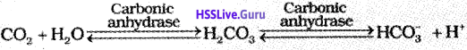
At the tissue site where partial pressure of CO2 is high due to catabolism, CO2 diffuses into blood (RBCs and plasma) and forms HCO2 and H+. At the alveolar site where pCO2 is low, the reaction proceeds in the opposite direction leading to the formation 0f CO2 and H2O. Every 100 ml of deoxygenated blood delivers approximately 4 ml of CO2 to the alveoli.