Kerala State Board New Syllabus Plus One Hindi Textbook Answers Unit 4 Chapter 13 अपराध Text Book Questions and Answers, Summary, Notes.
Kerala Plus One Hindi Textbook Answers Unit 4 Chapter 13 अपराध
प्रश्न 1.
छोटे भाई के प्रति बड़े भाई का लगाव सूचित करनेवाले वाक्य चुनें।
जैसेः खेल में अकेला होने पर भाई आकर मेरी मदद करता है।
उत्तर:
अकसर भाई मेरी वजह से ही हारते। फिर भी वे मुझसे कभी कुछ नहीं कहते थे।
प्रश्न 2.
निम्नलिखित चरित्रगत विशेषताओं के आधार पर तालिका भरें।
- पश्चातापग्रस्त
- दोस्ताना
- ईर्ष्यालु
- झूठा
- सहानुभूतिवाला
उत्तर:

प्रश्न 3.
‘वे मुझसे प्यार करते थे और मेरे प्रति उनका रुख एक संरक्षक की ज़िम्मेदारी जैसा था’ – ‘अपराध’ कहानी के आधार पर बड़े भाई की चरित्रगत विशेषताओं को विस्तार दें।
उत्तर:
सच्चा भाई
उदय प्रकाश की ‘अपराध’ कहानी के दो मुख्य कथापात्रों में से एक है बड़ा भाई। एक पैर को बचपन में पोलियो हो जाने से बड़ा भाई अपाहिज था। अपाहिज होने पर भी, खेल-खूदों में वह बड़ा तत्परता रखता था। बड़ा भाई अच्छा तैराक था। हाथ के पंजों की लड़ाई में वह बहुत निपुण था। खड़ब्बल जैसे खेलों में वह डूबा जाता था। खेल में विजय होते समय अतिप्रसन्न होना उसका स्वभाव था। छोटे भाई की ओर बड़े भाई के दिल में बड़ी हमदर्दी और वत्सलता थीं। बड़े भाई के चरित्र पर भाईचारे का गुण प्रकट करते हुए उदय प्रकाश जी लिखते हैं, छोटे भाई के प्रति उसका ‘रूख एक संरक्षक की जिम्मेदारी जैसा था”।
बड़े भाई के चरित्र पर दया, उदारता, सहायकस्वभाव आदि भी हम देखते हैं। वह बड़ा क्षमाशील था। भाईचारे में वह बड़ा ईमानदार था। शत्रुता मनोभाव उसके चरित्र में कभी भी हम नहीं देखते।
‘बहुत सुंदर थे, देवताओं की तरह…..’। उसकी सुन्दरता शरीर में ही नहीं, मन में भी था। त्याग, क्षमा, संयम, मौन-सहन आदि विशिष्ट गुणों से बड़े भाई का चरित्र अलंकृत है। दोस्ताना, सहानुभूति आदि चारित्रिक विशिष्टताओं से भी बड़ा भाई हमें आकर्षित करता है।
संक्षिप्त में हम इस प्रकार कहें बड़े भाई का चरित्र दूसरों में ईर्ष्या और आत्महीनता जगाने तक उदात्त और उत्कृष्ट था। छोटे भाई ने बड़े भाई के सामने अपनी अवस्था के बारे में खुद कहा है: “मैं ईर्ष्या, आत्महीनता, …… की आँच में झुलस रहा था।”
प्रश्न 4.
विवश छोटा भाई सालों बाद क्षमा माँगते हुए अपने बड़े भाई को पत्र लिखता है। वह पत्र लिखें।
(अथवा)
‘भाई ही मुझे क्षमा कर सकते हैं, जिन्हें मेरे झूठ का दंड भोगना पड़ा’। इस प्रसंग को लेकर लेखक, भाई को पत्र लिखता है। पत्र तैयार करें।
(अथवा)
‘तो इस अपराध के लिए मुझे क्षमा कौन कर सकता है’ – पश्चाताप से विवश छोटा भाई सालों बाद क्षमा माँगते हुए अपने बड़ा भाई को पत्र लिखता है। वह पत्र लिखें।
उत्तर:
शहडोल,
15.03.2016
प्रिय भाई,
आप कैसे हैं? कुशल में हैं न? आप जैसे एक भाई होना मेरा बड़ा सौभाग्य है।
भाई, मैं आप से एक बात साफ बताना चाहता हूँ। उस दिन मेरा खड़ब्बल चट्टान से टकराकर उछला और सीधे मेरे माथे पर आकर लगा। माथा फूट गया और खून बहने लगे। मैंने रोते हुए माँ को बताया कि मुझे आपने खडब्बल से मारा है। भाई! मुझे अच्छी याद है, इस पर आपको पिताजी से बहुत मार खाना पड़ा।
आज मैं पश्चाताप से विवश हूँ। आप के पैर पकड़कर क्षमा माँगता हूँ। मुझे क्षमा देंगे न? आप तो पहले ही बहुत अच्छे चरित्र के थे। आप शरीर और दिल दोनों में सुन्दर थे। मेरा अपराध क्षमा कीजिए….. कृपया क्षमा कीजिए…. आपको भागवान सदा संतुष्ट रखें……
(हस्ताक्षर)
आपका छोटा भाई
सेवा में,
जोन के.के.
सुन्दर घर,
बड़ा गाँव पी.ओ.
प्रश्न 5.
स्मृति में जब भी वे आँखें जाग उठती हैं, मेरी पूरी चेतना, ग्लानि, बेचैनी और अपराध-बोध से भर उठती है। छोटे भाई की प्रायश्चित भरी वाणी है। आवश्य ही आपको या आपके …….. को ऐसा कोई अनुभव हुआ होगा। उस अनुभव का वर्णन करें।
उत्तर:
जब मैं पाँचवीं कक्षा में पढ़ रहा था, तब मेरी जिंदगी में एक घटना हुई। उदय प्रकाश की ‘अपराध’ कहानी में वर्णित घटना के समान था वह घटना। ‘अपराध’ के कथापात्रों के समान माँ-बाप के लिए हम दो ही संतान थीं तब घर में। मैं और मेरा छोटा भाई। मैं स्वभाव से तेज़ था। कोपशील और स्वार्थ भी था। लालच भी था।
माँ ने एक दिन घर में बिरियाणी बनायी थी। खाने के समय के लिए बिरियाणी को माँ ने सुरक्षित रखा था। माँबाप खेत गये। इस अवसर का लाभ उठाकर मैंने बिरियाणी चोरी की। जब माँ वापस आयी, तब माँ को मालूम हुई कि बिरियाणी कम हो गयी है। माँ ने मुझे बुलाकर पूछा। लेकिन मैंने माँ से झूठ बोला कि छोटा भाई ने चोरी की है। बड़ी निपुणता से मैं ने माँ को समझाया कि मैं ने यह चोरी देखी है। मेरी बात को माँ ने विस्वास किया। छोटे भाई को पकड़कर माँ ने खूब मारा। छोटा भाई बड़ी आवाज में रोता था।
आज वर्ष अनेक बीत गये। छोटे भाई के विवाह का शुभदिन आ रहा है। मैंने यह निश्चय किया है कि शादी के पहले मैं अपने अपराध को छोटे भाई के सामने बताकर माफी माँगूगा।
Plus One Hindi अपराध Important Questions and Answers
प्रश्न 1.
मैं सबसे छोटा था और अकेला था। क्यों?
उत्तर:
उसका भाई और पूरे गाँव के सभी लड़के उससे छह वर्ष बड़े थे।
प्रश्न 2.
मुझे अपने भाई से ईर्ष्या होती थी, क्यों?
उत्तर:
भाई को बहुत सारे दोस्त थे।
प्रश्न 3.
भाई आकर मेरी मदद करते । कब?
उत्तर:
सब खेलते समय छोटा होने के कारण अकेला पड़ जाता तो, भाई आकर मदद करते थे।
प्रश्न 4.
दूसरे लड़के छोटे भाई को अपने पाली में शामिल क्यों नहीं करते थे?
उत्तर:
पाली में उसे शामिल करके हार का खतरा नहीं उठाना चाहते थे।
प्रश्न 5.
जोड़ी और पालीवाले खेलों में बड़े भाई क्या करते थे?
उत्तर:
बड़े भाई अपनी पाली में छोटे भाई को शामिल कर लेते थे।
प्रश्न 6.
“अकसर भाई मेरी वजह से ही हारते। फिर भी वे मुझसे कभी कुछ नहीं कहते थे.” क्यों?
उत्तर:
भाई के लिए लेखक एक उत्तरदायित्व की तरह था। लेखक को भाई बहुत प्यार करते थे और लेखक के प्रति भाई का रुख एक संरक्षक की जिम्मेदारी जैसा था। भाई यह जिम्मेदारी सदा निभाना चाहते थे।
प्रश्न 7.
शाम की धूप की विशेषता क्या है?
उत्तर:
शाम की धूप शरीर में उल्लास भरा करती है।
प्रश्न 8.
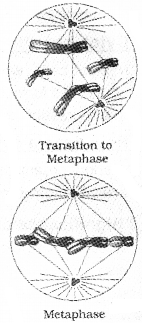
खडब्बल कैसे खेलता था?
उत्तर:
लकड़ी की छोटी-छोटी डंडियाँ हर लड़के के पास थीं। पूरी ताकत से खडब्बल को जमीन पर, आगे की ओर गति देते हुए, सीधे मारा जा रहा था।
प्रश्न 9.
छोटे भाई में किसकी ताकत न थी?
उत्तर:
छोटे भाई में इतनी ताकत न थी कि वह खड़बल को उतनी दूर तक पहुँचाता, जबकि वहाँ एक होड़, एक प्रतिद्वंद्धिता शुरु हो जाये।
प्रश्न 10.
गुस्से और तनाव में और ज्यादा ताकत से वे खड़ब्बल फेंक रहे थे-कौन?
उत्तर:
बड़ा भाई।
प्रश्न 11.
‘मुझे पहली बार यह लगा कि मैं वहाँ कहीं नहीं – क्यों?
उत्तर:
खड़ब्बल खेल में जीतते समय बड़ा भाई एक बार भी छोटे भाई की ओर नहीं देखता था। इतना ही नहीं, छोटे भाई को बड़ा भाई पूरी तरह उस समय भूलता था।
प्रश्न 12.
बड़े भाई के प्रति छोटे भाई के मन में कौन-सा भाव पैदा हुआ था?
उत्तर:
एक बहुत जबरदस्त प्रतिकार पैदा हो रहा था।
प्रश्न 13.
छोटा भाई किसकी आँच में झुलस रहा था?
उत्तर:
ईर्ष्या, आत्महीनता, उपेक्षा और नगण्याता की आँच में झुलस रहा था।
प्रश्न 14.
छोटे भाई के माथे पर कैसे चोट लगी?
उत्तर:
अचानक छोटे भाई का खड़ब्बल चट्टान से टकराकर उछला और सीधे उसके माथे पर आकर लगा। माथा फूट गया और खून बहने लगा।
प्रश्न 15.
बड़े भाई तेज़ी से दौड़ नहीं पा रहा था, क्यों?
उत्तर:
बड़े भाई का दायाँ पैर पोलियो का शिकार था।
प्रश्न 16.
घर पहुँचकर छोटे भाई ने माँ से क्या कहा?
उत्तर:
छोटे भाई ने माँ से यह कहा कि उसे बड़े भाई ने खड़ब्बल से मारा है।
प्रश्न 17.
बड़े भाई के प्रति छोटे भाई के मन में प्रतिकार की भावना क्यों उत्पन्न हुई?
उत्तर:
छोटे भाई के मन में ऐसा लग रहा था कि बड़े भाई के सामने वह कहीं नहीं है। बड़े भाई से इस प्रकार की उपेक्षा का अनुभव महसूस करने के कारण उसके दिल में बड़े भाई के प्रति प्रतिकार की भावना उत्पन्न हुई।
प्रश्न 18.
बड़े भाई की आँखों में करुणा और कातरता थीं- क्यों?
उत्तर:
बड़े भाई के विरुद्ध छोटा भाई झूठ बोल देने पर बड़े भाई को पिताजी से पीट सहना पड़ा। इसलिए बड़े भाई की आँखों में करुणा और कातरता थीं।
प्रश्न 19.
छोटे भाई के मन में जब बचपन की उस घटना की स्मृतियाँ आती हैं तब उसे कैसा अनुभव होने लगता है?
उत्तर:
छोटे भाई की पूरी चेतना ग्लानि, बेचैनी और अपराध बोध से भर आती है।
प्रश्न 20.
वे इस घटना को पूरी तरह भूल चुके हैं – कौन?
उत्तर:
बडा भाई।
प्रश्न 21.
छोटे भाई ने अपने अपराध के लिए क्षमा माँगनी चाही, लेकिन असफल रहा – क्यों?
उत्तर:
माँ-बाप मर गये थे। बातों की सत्यावस्था ठीक-ठीक उन्हें समझाने के लिए अब अवसर नहीं। इतना ही नहीं बड़ा भाई इन पूरी बातों को भूल गया है।
प्रश्न 22.
अब यह निर्णय बदला नहीं जा सकता -क्यों?
उत्तर:
छोटे भाई के झूठ का दंड बड़े भाई को भोगना पड़ा। लेकिन बड़ा भाई यह घटना बिलकुल भूल चुके थे। उस समय झूठ बोलने का जो निर्णय लिया था वह गलत और अन्यायपूर्ण था। लेकिन वह निर्णय बदलना अब संभव नहीं।
प्रश्न 23.
बड़े भाई के विरुद्ध छोटे भाई से हुए अपराध के बारे में बताकर अपने मित्र के नाम पत्र लिखता है। वह पत्र तैयार कीजिए।
उत्तर:
स्थान,
तारीख,
प्रिय मित्र,
मेरी बात जानकर तुम असंतुष्ट हो जाओगे कि यह कितनी पुरानी बात है! लेकिन मेरे मन में यह अब भी एक काँटा जैसी है वह बात।
बात यह है कि बचपन में एक दिन मेरा भाई मुझे भूलकर खड़ब्बल खेल रहा था। छोटा होने के कारण अपनी पाली में उस दिन उन्होंने मुझे शामिल नहीं किया था। जीतने की खुशी में भाई ने मेरी और देखा तक नहीं। इससे में रो पड़ा। मैं अकेले खड़ब्बल को पत्थर पर फेंककर खेलते वक्त अचानक वह मेरे माथे पर लगा। भाई मेरे पास दौडकर आये। लेकिन मैं ने उसको रोका | माँ से मैंने झूठ कह दिया कि भाई ने मुझे मारा है। पिताजी ने उन्हें इस पर खूब पीटा। यह सज़ा मिलते समय भाई ने मुझपर करुणा भरी दृष्टि से देखा। भाई का वह कारुणिक अवस्था मेरी स्मृति में अब भी है। मैं उस गलती केलिए क्षमा माँगना चाहता हूँ। लेकिन कैसे? माँ-बाप मर गये। भाई यह बात भूल गया है। मेरा मन अशांत है। मित्र, तुम इसकेलिए एक परिहार बता दो।
मेरा विश्वास है कि तुम जवाब ज़रूर दोगे।
प्यार से,
(हस्ताक्षर)
नाम
प्रश्न 24.
कहानी का अंश पढ़ें और प्रश्नों के उत्तर लिखें।
मैंने भाई का चेहरा देखा। वे मेरी ओर देख रहे थे। उनकी आँखें लाल थीं और उनमें करुणा और कातरता थीं, जैसे वे मुझसे याचना कर रहे हों कि में सच बोल दूं। लेकिन तब तक देर हो चुकी थी। उन्हें सज़ा मिल चुकी थी। फिर इतनी जल्द बात को बिलकुल बदलना मुझे संभव भी नहीं लग रहा था। क्या पता, पिताजी फिर मुझे ही मारने लगते। में डर रहा था।
i. यह किस कहानी का अंश है?
उत्तर:
उदय प्रकाश की ‘अपराध’ कहानी का।
ii. बड़े भई की आँखों में कौन-सा भाव था?
उत्तर:
करुणा और कातरता थीं।
iii. छोटा भाई क्यों डर रहा था?
उत्तर:
छोटे भाई ने जो बात बताई थी उसे जल्द बिलकुल बदलना उसे संभव नहीं लग रहा था। इसलिए पिताजी सच जानते समय छोटे भाई को भी पिताजी से मार मिलने की संभावना थी। इन बातों से छोटा भाई डर रहा था।
iv. उस दिन के छोटे भाई की डायरी लिखें।
उत्तर:
25 मार्च 2016
शहडोल :
आज मेरे लिए बिलकुल बुरा दिन था। मैंने झूठ बोलकर बड़े भाई को पिताजी के सामने अपराधी बनाया। बेचारा भाई! उनकी आँखें उस समय लाल हो गयी थीं। उनमें करुणा और कातरता थीं। वे मुझसे मौन याचना कर रे थे कि मैं पिताजी से सच बोल दूं। लेकिन मुझसे वह नहीं कर पाया। बड़े भाई को पिताजी से मेरे झूठ से सजा मिल गया। मैं विवश हो गया था। जल्द बात को बिलकुल बदलना मुझे संभव भी नहीं लग रहा था। मुझे यह डर भी था कि पिताजी मुझे सच जानते समय बुरी तरह मारेंगे।
हे भगवान! मुझसे बड़ा अपराध हो गया। मुझे क्षमा कर । भाई को अनुग्रह दे। मुझे नींद नहीं आती। मेरा मन बहुत व्याकुल है।
प्रश्न 25.
वह एक धीर, निडर एवं साहसी सैनिक था। अपने देश की सेना का नेतृत्व करनेवाला सेनानायक। उन दिनों दुश्मन सेना के साथ उनका युद्ध चल रहा था। दोनों सेनाओं में काफी विनाश हो चुका था। फिर भी सेनानायक निडर होकर अपनी सेना का नेतृत्व कर रहा था। हर दिन युद्ध समाप्त होने के बाद सब अपने-अपने डेरे में जाया करते थे। सेनानायक का निर्देश था कि कोई उसे तंग न करे क्योंकि शाम को वह भगवान की पूजा करेगा। रात को सैनिकों ने देखा कि सेनानायक कहीं जा रहा है। मैनिकों ने चुपचाप उनका पीछा किया। उन्होंने देखा कि सेनानायक युद्ध के मैदान में घायल पड़े सैनिकों की सेवा कर रहा है।, घावों में पट्टी बाँध रहा है और दवा लगा रहा है। आश्चर्य की बात यह थी कि उनमें शत्रु पक्ष के घायल सैनिक भी थे। सैनिकों के पूछने पर सेनानायक ने कहा कि घायलों की सेवा करते समय शत्रु और मित्र का भेद-भाव न दिखाएँ। इनकी सेवा ही मेरे लिए भगवान की पूजा है।
i. सेनानायक रोज़ शाम को कहाँ जा रहा था?
उत्तर:
सेनानायक युद्ध के मैदान में घायल पड़े सैनिकों की सेवा कर रहा था, घावों में पट्टी बाँध रहा था और दवा लगा रहा था। आश्चर्य की बात यह थी कि उनमें शत्रु पक्ष के घायल सैनिक भी थे।
ii. सेनानायक की चरित्रगत विशेषताओं पर प्रकाश डालें।
उत्तर:
साहसी सैनिक
सैनिक में बड़ा नेतृत्व गुण था। इसलिए वे सेनानायक बन गये। वे बड़े बुद्धिमान थे। शत्रुओं को पराजित करने के लिए उन्होंने बुद्धि से काम किया। निर्भयत्व उसके चरित्र का एक विशिष्ट गुण था। सेनानायक वीर-शूर होने पर भी बड़ी सेवा मनोभाव से जीनेवाले भी थे। वे आदर्श सैनिक थे। एक आदर्श सैनिक शत्रुसेना के सामने भी मर्यादा और सेवाभाव से व्यवहार करता है। सेनानायक की रोगी – शुश्रूषा से हमें ज्ञात होते हैं कि उनका चरित्र आदर्श और मर्यादा से अलंकृत था।
प्रश्न 26.
यह घटना पढ़ें और प्रश्नों के उत्तर लिखें।
शादी में कार मिली। पत्नी ने अपने पति से कहाः सुनो, हम भी एक ड्राइवर रख लें? तुम्हारे घर में कई कारें थीं, तुमने कार चलाना क्यों नहीं सीखा? कई कारें थीं तो कई ड्राइवर भी थे….. कार चलाना सीखने की कभी ज़रूरत भी महसूस नहीं हुई….फिर भी मैंने सीखनी चाही थी, पर पापा-मम्मी ने नहीं सीखने दी। क्यों? भाई ने सीखी थी। एक बार उसका एक्सीडेंट हो गया… यह समझो कि वह मरते मरते बचा था, तब से उसकी ड्राइविंग पर पाबंदी लगा दी गई थी और मुझे भी सीखने से मना कर दिया। एक्सीडेंट क्या ड्राइवर से नहीं हो सकता? हो सकता है किंतु खतरा अक्सर आगे बैठनेवाले का ही होता है, हाध-पैर टूटेंगे या मरेगा तो ड्राइवर मरेगा।
i. शादी में क्या मिली?
उत्तर:
शादी में कार मिली।
ii. एक्सीडेंट में खतरा अक्सर किसका होता है?
उत्तर:
आगे बैठनेवाले का ही होता है।
iii. पत्नी ने कार चलाना क्यों नहीं सीखा?
उत्तर:
कार चलाते समय पत्नी के भाई को बुरी तरह एक्सीडेंट हो गयी थी। वह मरते – मरते बचा था। तब से पापामम्मी ने भाई की ड्राइविंग पर पाबंदी लगा दी गयी और पत्नी को भी सीखने से मना कर दिया।
iv. उपर्युक्त घटना का संक्षेपण करें।
उत्तर:
शादी में मिली कार एक्सीडेंट की डर से पत्नी नहीं चलाती है। पति से पत्नी ड्राइवर को रखने का आग्रह प्रकट करती है। लेकिन पत्नी को पति समझाता है कि ड्राइवर को भी एक्सीडेंट हो सकता है।
v. संक्षेपण के लिए शीर्षक लिखें।
उत्तर:
ड्राइविंग और एक्सीडेंट।
vi. मान लें, शादी में पत्नी को नई मॉडल कार मिली है। उस पॉडल कार की बिक्री बढ़ाने केलिए अखबार में छपे कंपनी का विज्ञापन तैयार करें।
उत्तर:

अपराध Previous Years Questions & Answers
प्रश्न 1.
‘अपराध’ कहानी का ये वाक्य पढ़िए।
“मैं अपने इस अपराध के लिए क्षमा माँगना चाहता हूँ।
इस अपराध की सज़ा पाना चाहता हूँ।”
अपने अपाहिज भाई का पिताजी से सज़ा दिलाते हुए उस दिन की छोटे भाई की डायरी तैयार कीजिए।
- भाई का प्यार
- खडब्बल के खेल में भाई की जीत
- उपेक्षा की तीव्र वेदना
- माँ बाप से झूठ बोलना
उत्तर:

अपराध Summary in Malayalam


अपराध शब्दार्थ